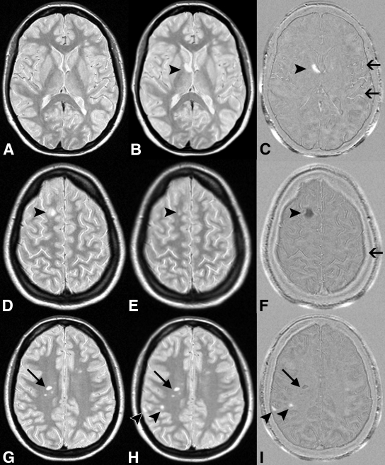
Figure 1:
MS activity on, A, D, G, halfway-registered baseline PD-weighted MR images; B, E, H, halfway-registered follow-up PD-weighted images; and C, F, I, subtraction images. B, C, Arrowheads = new lesion (positive activity) shown as hyperintensity on C. Arrows = flow artifacts. D, E, F, Arrowheads = shrunken lesion (negative activity) shown as hypointensity on F. Arrow = residual registration artifact. G, H, I, Arrowheads = new lesions, one of which is juxtacortically located and easily missed on H but is clearly visible on I. Arrows = false-positive negative activity. Lesion was visible on both G and H and did not decrease in size.

