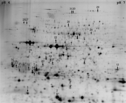
Fig. 1.
Preparative 2D gel showing the manually excised spots for MS/MS analysis. The pH 4–7 IPG strip was loaded with 400 μg of proteins resolved in the second dimension using a 12.5% SDS–polyacrylamide gel. The gel was then stained with Sypro Ruby. A manual matching was performed with DIGE analytical gels in order to locate the differentially expressed proteins. Protein identifications are presented in Supplementary Tables S1 and S2 at JXB online. The evolution of standard abundances of proteins are presented in Tables 2, 3, and Supplementary Table S3 at JXB online.

