Figure 3.
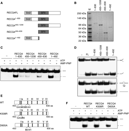
Domain analyses of the RECQ4 protein. (A) Schematic diagram of the RECQ4 fragments. The conserved SFII helicase domain is shown in dark grey, whereas the Sld2-like domain is shown in light grey. (B) Visualization of recombinant RECQ4 fragments purified from E. coli by SDS–PAGE and Commassie blue staining. (C) Helicase activities of the RECQ4 fragments (20 nM each) using duplex DNA in the presence of ATP or AMP-PNP. (D) Helicase activities of the RECQ4 fragments (20 nM each) using splayed arm (upper panel) or 12-nt bubble (lower panel). (E) Schematic diagram of the RECQ4 SFII helicase domain with the seven conserved helicase motifs. Walker A motif (GAGKS) and DEAH box belonging to the Walker B motif are shown. Walker A mutant K508R has Lys508 mutated to Arg508, whereas Walker B mutant D605A has Asp605 mutated to Ala605. (F) Helicase activities of the full-length RECQ4 WT, K508R and D605A proteins (20 nM each) using duplex DNA in the presence of ATP or AMP-PNP.

