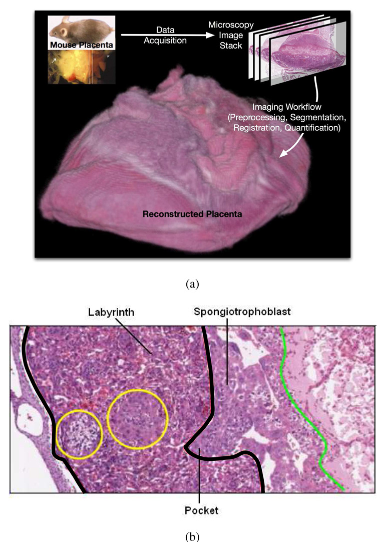
Figure 1.
(a) A mouse placenta reconstructed in 3D with the described imaging workflow. (b) Zoomed placenta image showing the different tissue layers. The tissue between the two thick black boundaries is the labyrinth tissue. The pocket area is an example of the infiltration (interdigitation) from the spongiotrophoblast layer to the labyrinth layer. The cells in the left circle are glycogen cells.

