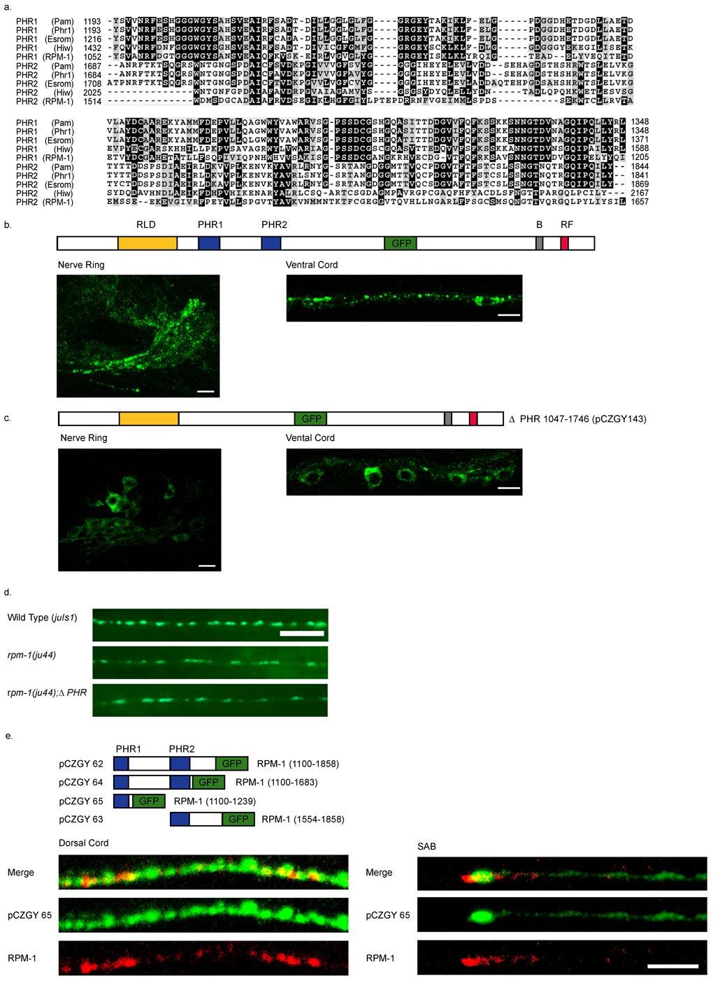
Figure 6. The PHR domain of RPM-1 is necessary and sufficient for synaptic localization.
(a) A CLUSTAL alignment of the PHR regions. (b–c) Confocal images of GFP fluorescence in transgenic animals. (b) Expression of the functional full-length RPM-1::GFP(juIs58) is primarily in the nerve ring and nerve cords, and barely detectable in the cell bodies. (c) Deletion of the PHR domains juEx143 [pCZGY143] causes GFP to be confined to the cell bodies and is barely seen in nerve processes. (d) Expression of RPM-1ΔPHR::GFP (pCZGY143) does not rescue rpm-1(ju44). Wild type animals expressing juIs1[P unc-25 SNB-1::GFP] show an array of evenly sized, evenly spaced fluorescent puncta (top). rpm-1(ju44) animals display enlarged puncta with gaps in SNB-1GFP expression (middle), and are not rescued by the expression of RPM-1ΔPHR::GFP (pCZGY143) (bottom). (e) Schematics of the constructs expressing regions of PHR fused to GFP. The images are juEx1172 [pCZGY65] (pan neural PHR1::GFP) co-stained with endogenous RPM-1 and GFP in the dorsal cord (left panels) and in the SAB neurons (right panels). Scale bars 5µm.

