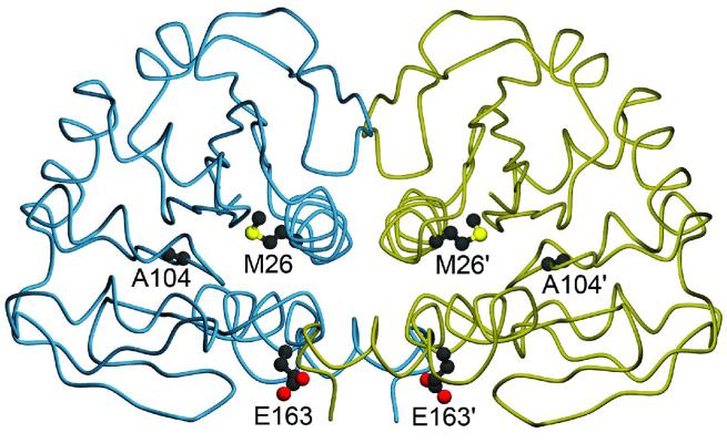
Figure 1.
Location of three PD-associated mutations in the DJ-1 dimer. The DJ-1 dimer is represented with one monomer colored blue and the other gold. Three residues that are mutated in certain forms of familial Parkinsonism studied in this work are shown in both monomers of the DJ-1 dimer, with prime symbols indicating the symmetry-related residues. Both M26 and A104 are located in the core of dimeric DJ-1, while E163 is a surface-exposed residue in α-helix G. The figure was created with POVscript+(65).

