
Figure 2.
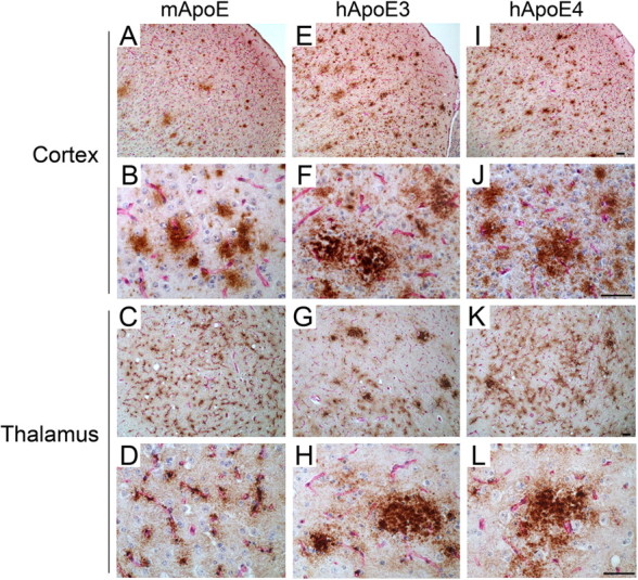
Human ApoE3 and ApoE4 alter the Aβ deposition pattern in Tg-SwDI mice. Brain sections from 12-month-old mice were double immunostained for Aβ (brown) and collagen type IV (red) to identify cerebral microvessels. A–L, Tg-SwDI/muAPOE mouse cortex (A, B) and thalamus (C, D); Tg-SwDI/hAPOE3/3 mouse cortex (E, F) and thalamus (G, H); Tg-SwDI/hAPOE4/4 mouse cortex (I, J) and thalamus (K, L). Note that the prevalent thalamic microvascular-associated Aβ deposition in Tg-SwDI/muAPOE mice (C, D) was markedly reduced in the Tg-SwDI/hAPOE3/3 mice (G, H) and Tg-SwDI/hAPOE4/4 mice (K, L). Scale bars, 50 μm.