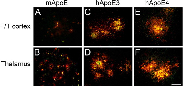
Figure 6.
Colocalization of ApoE proteins and fibrillar amyloid deposits in Tg-SwDI mouse brain. Brain sections from 12-month-old mice were labeled for fibrillar Aβ using thioflavin-S (green) and ApoE (red). A–F, Tg-SwDI/muAPOE mouse cortex (A) and thalamus (B); Tg-SwDI/hAPOE3/3 mouse cortex (C) and thalamus (D); Tg-SwDI/hAPOE4/4 mouse cortex (E) and thalamus (F). Note the strong colocalization of mouse ApoE with microvascular amyloid deposits in Tg-SwDI/muAPOE mice and human ApoE with parenchymal amyloid plaques in Tg-SwDI/hAPOE3/3 and Tg-SwDI/hAPOE4/4 mice. Scale bar, 50 μm. F/T, Frontotemporal cortex.

