Figure 8.
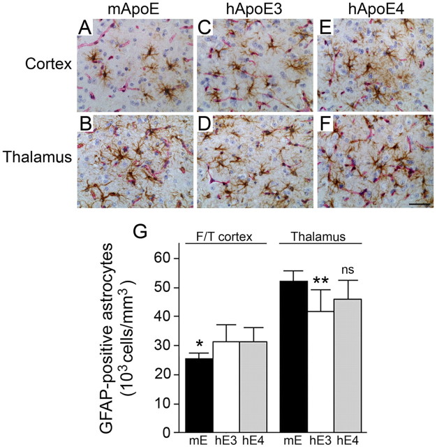
Human ApoE3 and ApoE4 increase cortical and decrease thalamic reactive astrocyte numbers in Tg-SwDI mice. Brain sections from 12-month-old mice were double immunostained with GFAP to reveal astrocytes (brown) and collagen type IV (red) to identify cerebral microvessels. A–F, Tg-SwDI/muAPOE mouse cortex (A) and thalamus (B); Tg-SwDI/hAPOE3/3 mouse cortex (C) and thalamus (D); Tg-SwDI/hAPOE4/4 mouse cortex (E) and thalamus (F). Scale bar, 50 μm. G, Quantitative stereological estimation of astrocyte densities showed increased numbers in the frontotemporal cortex and decreased numbers in the thalamus of Tg-SwDI/APOE3/3 mice (white bars) and Tg-SwDI/APOE4/4 mice (gray bars) compared with Tg-SwDI/muAPOE mice (black bars). The data presented are the mean ± SD (n = 8 animals per group). In the cortex, Tg-SwDI/muAPOE mice had lower numbers of astrocytes compared with Tg-SwDI/hAPOE3/3 mice or Tg-SwDI/hAPOE4/4 mice (*p < 0.02). In the thalamus, Tg-SwDI/muAPOE mice had higher numbers of astrocytes compared with Tg-SwDI/hAPOE3/3 mice (**p < 0.01) and Tg-SwDI/hAPOE4/4 mice (p < 0.06, not significant). F/T, Frontotemporal cortex.

