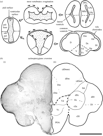
Figure 1.
(a) Schematic of telencephalic development of actinopterygians (teleost as an example) in comparison with other vertebrates and (b) (i) a frontal section of the telencephalon of tilapia Oreochromis niloticus stained by cresyl violet and (ii) line drawing indicating telencephalic structures. Figure 1a adapted from Yamamoto et al. (2007). Thick broken lines in (a) and large dots in (b) indicate the boundary between the areas dorsalis (D: presumed pallial homologue) and ventralis (V: presumed subpallial homologue). ca, commissura anterior; dDc, dorsal region of Dc; dDm, dorsal region of Dm; dDml, lateral portion of dDm; dDmm, medial portion of dDm; DPa, dorsal pallium; LPa, lateral pallium; MPa, medial pallium; POA, area preoptica; Se, septum; St, striatum; vDc, ventral region of Dc; vDm, ventral region of Dm; VPa, ventral pallium. See text for other abbreviations. Scale bar, 1 mm.

