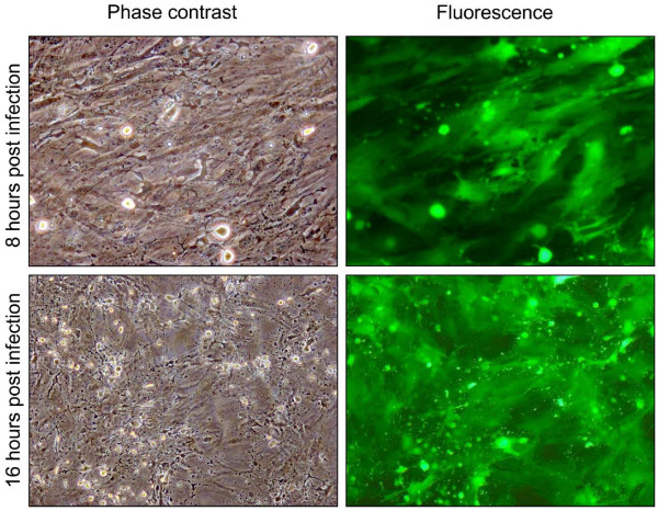
Figure 2.
Representative phase contrast images and fluorescence (green) (10×) of bovine endometrial stromal cells at different time (12, 24, 48, 72 h) post infection (P.I.) with 1 m.o.i. of BoHV-4 EGFPΔTK and the respective phase contrast images of uninfected control. Spreading of the infection can be observed by the green colour invading the field during the time and the CPE is morphologically appreciable by the change of the cell shape, where the cells tend to shrink, becoming roundest and detaching the flask surface. The experiment was repeated three times giving the same result. Each experiment was repeated three times giving similar results.

