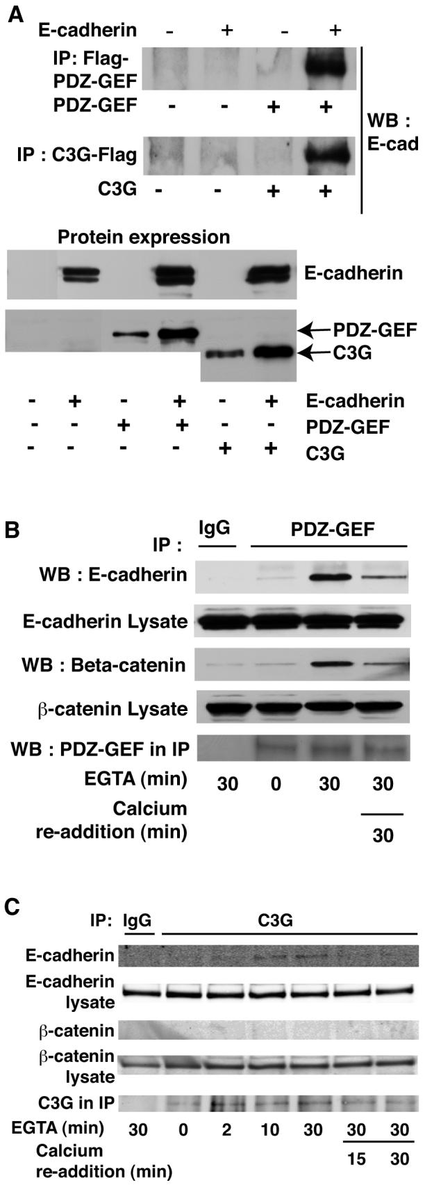
Fig. 5. E-cadherin co-precipitates with PDZ-GEF I and C3G.

A: E-cadherin was exogenously expressed in 293T cells along with either Flag tagged PDZ-GEF I or C3G. Flag antibody was used to precipitate GEFs prior to blotting for E-cadherin. E-cadherin co-precipitated with both PDZ-GEF I (top panel) and C3G (middle panel). Expression of proteins is shown below. B: Endogenous E-cadherin and β-catenin associate with PDZ-GEF I in MDCK cells. MDCK cells were subject to calcium switch and PDZ-GEF antibody used to immunoprecipitate the endogenous GEF. Association of endogenous E-cadherin with PDZ-GEF was greatly enhanced upon disruption of junctions with EGTA for 30 min but returned to near basal levels following Ca2+ re-addition. β-Catenin also coprecipitated with the E-cadherin and PDZ-GEF I complex (lower panels). C: E-cadherin but not β-catenin co-immunoprecipitated with C3G in MDCK cells. MDCK cells were calcium switched as above and C3G antibody used for immunoprecipitation. Data representative of at least three independent experiments.
