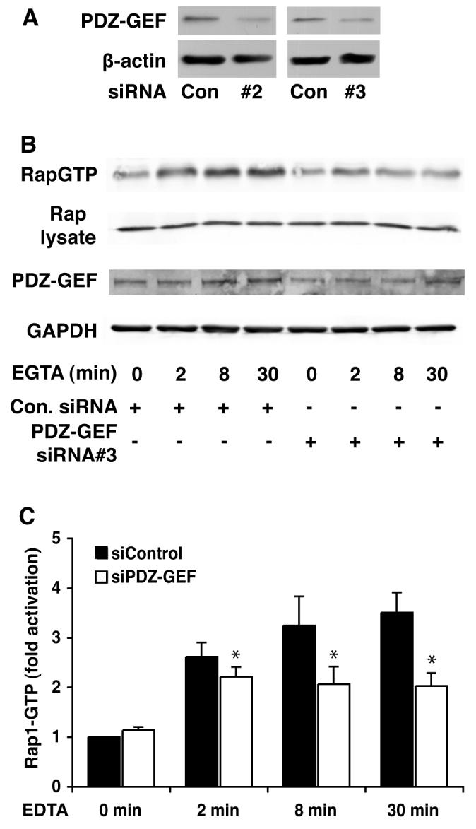
Fig. 6. Knockdown of MDCK cell PDZ-GEF I reduces the ability of cell junction disruption to activate Rap1.

A: Transfection of two different PDZGEF siRNAs resulted in decreased expression of the GEF 48 hr later. B: MDCK cells were transfected with control or two different PDZ-GEF I siRNAs (75 mM siRNA#3 shown). Forty-eight hours later calcium switch was performed as indicated and RapGTP levels measured using the RalGDS-RBD pull down. Glyceraldehyde 3 phosphate dehydrogenease and total Rap1 expression levels indicate equal loading. C: Graph shows quantitation of the data represented in (B) (mean +/− SEM from three separate experiments using siRNA #3).
