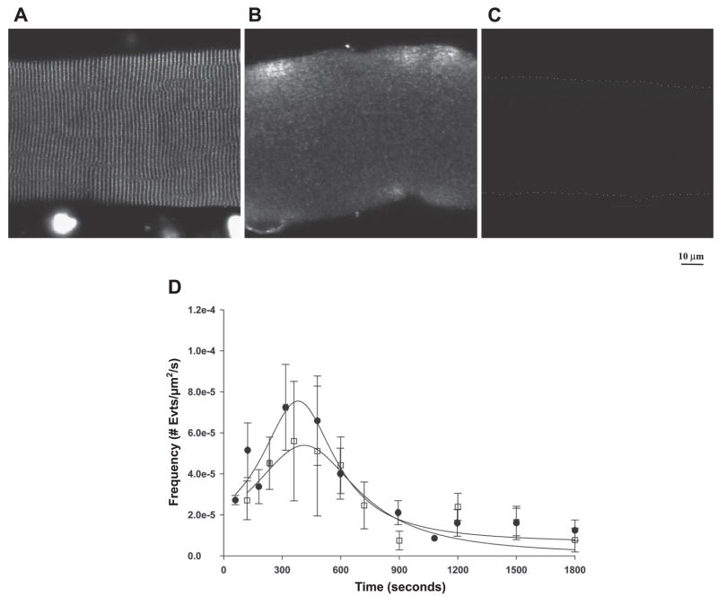
Fig. 8.
Effect of a myosin light chain kinase (MLCK) CaM binding peptide on localization of CaM and the time-dependent appearance of Ca2+ sparks in diaphragm. Immunofluorescence localization of endogenous CaM shows that the MLCK CaM binding peptide (10 μM) results in a loss of endogenous CaM (A and B). C: no detectable fluorescence from fibers incubated in secondary antibody alone is shown. Dotted line outlines the cell for visual purposes. D: no significant alteration in the appearance of Ca2+ sparks was observed in fibers incubated with the MLCK CaM binding peptide (squares) and controls (circles).

