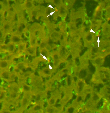
Figure 5:
Immunofluorescence staining of liver tissue for MBs (arrows) and Kupffer cells (arrowheads). Targeted MBs were located within Kupffer cells as soon as 4 minutes after intravenous administration. For MB staining, slices were incubated with Texas red–conjugated rabbit antistreptavidin primary antibody (red). For Kupffer cell staining, slices were incubated with primary monoclonal rat antimouse F4/80 antibody and secondary fluorescein isothiocyanate–labeled polyclonal goat antirat IgG2b antibody (green). (Original magnification, ×400.)

