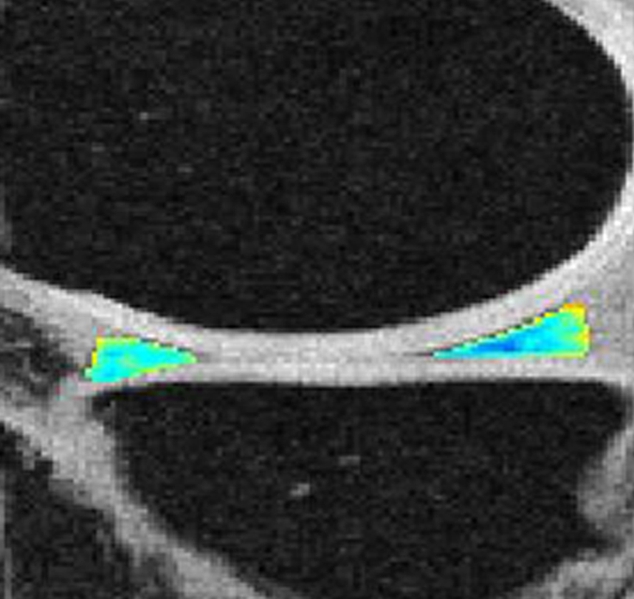
Figure 2h:

Representative MR images show medial meniscus in (a, d, g) a healthy subject, (b, e, h) a patient with mild OA, and (c, f, i) a patient with severe OA. (a–c) Fast spin-echo images (4300/51) show morphology of the menisci. In a and b, the meniscus appears normal, but c shows a tear in the anterior and posterior horns of the medial meniscus. Corresponding (d–f) T1ρ (in milliseconds) and (g–i) T2 (in milliseconds) color maps overlaid on SPGR images (20/7.5, 12° flip angle) clearly show differences in the meniscal matrix among the three subjects.
