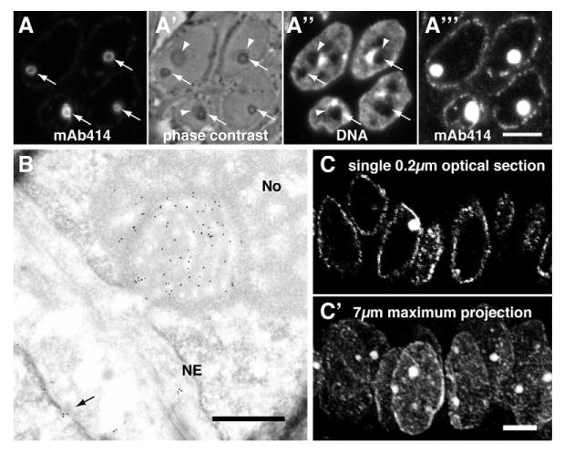
Fig. 1.

The monoclonal antibody 414 (mAb414) directed against nuclear pore complex (NPC) proteins exhibits a strong preference for NCSs. (A) Double fluorescence of mAb414 (A) and DAPI DNA stain (A″) on a semi-thin frozen section of human endometrium in the secretory phase. NCS fluorescence appears as rings (A, arrows). The rings, i.e. the matrix and membrane tubules of NCSs, appear as phase-dense circles in phase contrast microscopy (A′, arrows). Moreover, NCSs are often encircled by nucleoli (arrowheads) and, like nucleoli, appear chromatin-free (A″). The concentration of mAb414 antigens in NCSs is so high that the classical rim staining of NPCs only becomes visible if the image is overexposed to an extent that saturates NCS staining (A‴). (B) MAb414 immunogold-stained electron micrograph of an ultrathin cryosection of luteal human endometrium. Note the strong and specific gold labeling of a grazing section of a NCS (i.e. its core is covered by its membrane tubules and matrix) that is embedded in a nucleolus (No) and attached to the nuclear envelope (NE). At least one NPC of a neighboring cell is identified by mAb414 (arrow). (C) Confocal micrograph of indirect mAb414 fluorescence of a 7-μm-thick paraffin section of luteal human endometrium. In a single 0.2 μm optical section, a NCS is visible in only one of the nuclei defined by the classical rim staining of NPCs (C), whereas, in a maximum projection of all optical planes, all nuclei outlined by hazy NPC staining contain NCSs (C′). Scale bars: 5 μm.
