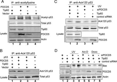
Figure 5.
PDCD5 promotes p53 acetylation and p53-dependent apoptosis-related gene expression mediated by Tip60. (A) U2OS cells were cotransfected with Tip60 and either PDCD5 or empty vector for 36 hours. After 4 hours of treatment with 10 mM sodium butyrate, total cell lysates were subjected to IPs with a pan-acetyl-lysine antibody. Precipitates were blotted with an anti-p53 antibody to detect the p53 acetylation level. Input lysates were also blotted with anti-p53, anti-Tip60, anti-PDCD5, and antiactin antibodies. (B) H1299 cells were cotransfected as indicated for 36 hours. After 4 hours of treatment with 10 mM sodium butyrate, total cell lysates were subjected to IPs with an anti-Ack 120-p53 antibody. Precipitates were blotted with an anti-p53 antibody to detect K120 acetylation of p53. Input lysates were also blotted with anti-p53 antibodies. (C) U2OS cells were transfected as indicated. After 24 hours, cells were given UV irradiation for 12 hours. Immunoprecipitation and Western blot were performed as in (B). (D) U2OS cells were transiently cotransfected with Tip60 and either siPDCD5 or control siRNA. After 24 hours, cells were treated with or without genotoxic treatments as indicated, and then total cell lysates were analyzed for the presence of Bax, PDCD5, and actin by Western blot.

