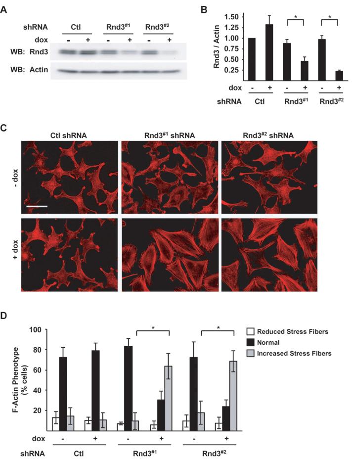
Figure 1.

Depletion of endogenous Rnd3 induces melanoma actin cytoskeletal reorganization. Doxycycline-inducible melanoma cells expressing Ctl, Rnd3#1 or Rnd3#2 shRNA. A, Cell lysates analyzed by western blotting for Rnd3 and β-actin protein levels. B, Quantitation of endogenous Rnd3 knockdown. Graphed are the means ± SD Rnd3/actin ratios from three experiments with the Ctl shRNA condition set to one. Asterisk denotes statistical significance as determined by a two-tailed unpaired t-Test comparing Rnd3 knockdowns with Ctl knockdowns (* P<0.05). C-D, F-actin organization in inducible shRNA melanoma cells. C, Cells cultured ± dox for 72 hours were fixed and stained with TRITC-phalloidin. Bar, 50 μm. D, Quantitation of the F-actin phenotype shown in (C). The graph displays the proportion of cells with visibly altered actin stress fiber organization; means ± SD of >600 cells from multiple independent experiments; (*P<0.05).
