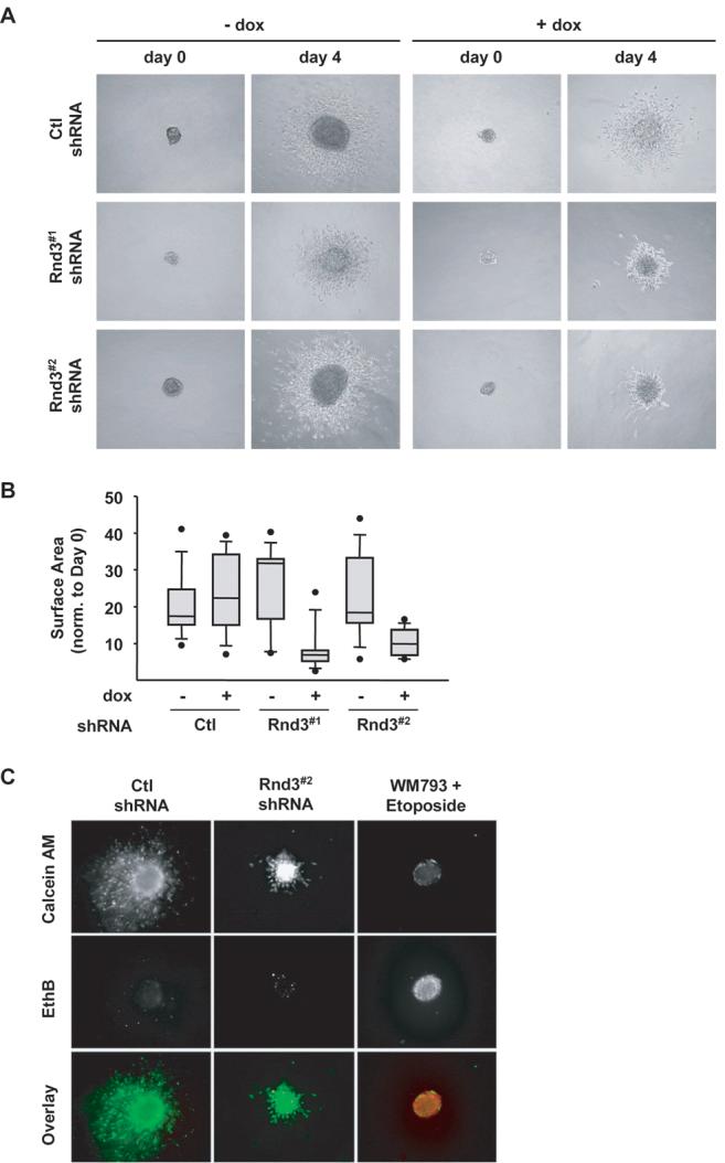
Figure 5.

Melanoma invasive outgrowth is inhibited following Rnd3 knockdown. Spheroids formed from Ctl, Rnd3#1 and Rnd3#2 shRNA WM793TR cells embedded in a 3-D collagen gel for 4 days. A, Phase contrast pictures displaying spheroid morphology at day 0 and day 4 in ± dox. B, Quantitation of spheroid outgrowth shown in (A). Spheroid surface area measured at day 4 normalized to its area at day 0. C, Cell viability within spheroids after 4 days embedded in a collagen gel, live cells stain green (calcein-AM) and dead cells stain red (ethidium bromide).
