Figure 6.

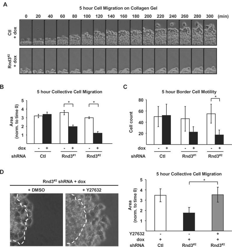
Rnd3 expression is required for directed melanoma cell migration on 3-D collagen gels. Spheroids from Ctl, Rnd3#1 and Rnd3#2 shRNA cells cultured in complete medium ± dox on top of a collagen gel. A, Micrographs of spheroids acquired from time lapse microscopy over a five hour imaging period. B-C, Quantitation of spheroid migration depicted in (A). B, Collective cell movement, measured as the increase in spheroid surface area over time (* P<0.05). C, The number of border cells, determined by counting individual cells separated from the cell sheet (* P<0.05). D, Collective cell movement of Ctl and Rnd3#2 shRNA cells treated ± 5 μM Y27632 one hour prior to plating on collagen gels. Images show an overlay of the spheroid surface area at time 0 (outlined by white line) superimposed onto its area five hours later. Graph depicting quantitation of the collective cell movement in spheroids treated ± Y27632 (* P<0.05).
