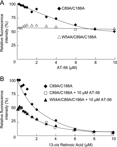
FIGURE 4.
Tryptophan fluorescence quenching by AT-56. A, fluorescence quenching of intrinsic tryptophan of C89A/C186A mutant (♦) and W54A/C89A/C186A mutant (▴) of mouse L-PGDS by incubation with AT-56. B, fluorescence quenching of intrinsic tryptophan of the mouse L-PGDS C89A/C186A mutant in the absence (♦) or presence (□) of 10 μm AT-56 and that for the W54A/C89A/C186A mutant in the presence of 10 μm AT-56 (▴) by incubation with 13-cis-retinoic acid.

