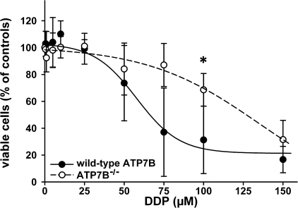
FIGURE 2.
Viability of primary hepatocytes from wild-type (•) and Atp7b-/- (○) livers after treatment with DDP. Cell survival was measured by MTT assay. Control cells were cultured without DDP treatment. The data show the means ± S.D. of four separate experiments performed in triplicate and presented as percentages of untreated control. The difference at a concentration of 100 μm DDP was significant (*, p < 0.05). The curves were fitted using a sigmoidal decay model.

