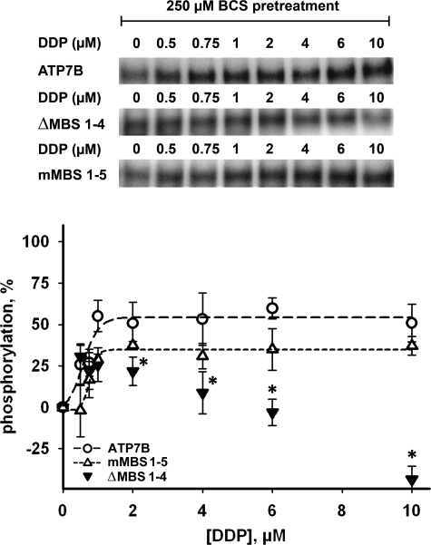
FIGURE 7.
The effects of DDP on catalytic phosphorylation of ATP7B, ΔMBS1–4 and mMBS1–5. Wild-type ATP7B (○), ΔMBS1–4 (▾), and mMBS1–5 (▵) were inactivated by pretreatment with 250 μm BCS, incubated with DDP (0.5–10 μm) for 10 min and then phosphorylated with [γ-32P]ATP. The phosphorylation levels of ATP7B in the presence of BCS were subtracted, and the maximum phosphorylation level induced by copper was set to 100%. In the upper panel a typical autoradiogram is shown. The lower panel shows quantification (means ± S.D. of three separate experiments). The curves for ATP7B and the mMBS1–5 mutant were fitted with a sigmoidal model. The data for ΔMBS1–4 could not be fitted using the sigmoidal model and are shown without fitting. *, p < 0.05, when comparing ΔMBS1–4 with wild-type ATP7B under DDP treatment.

