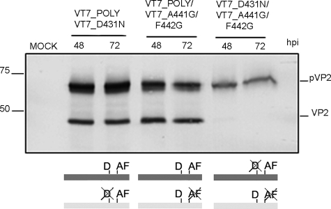
FIGURE 7.
pVP2 intramolecular proteolytic processing. Western blot analysis of proteins expressed in QM7 cells coinfected with VT7LacOI/POLY and VT7LacOI/D431N, VT7LacOI/POLY and VT7LacOI/A441G/F442G, or VT7LacOI/A441G/F442G and VT7LacOI/D431N. Infected cultures were harvested at 48 and 72 h pi (hpi). A scheme indicating the combinations of rVV used in this experiment (mutated positions are crossed out) is shown at the bottom.

