FIGURE 4.
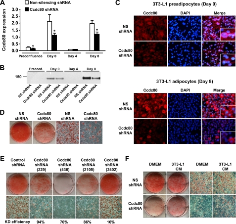
Knockdown of Ccdc80 inhibits adipocyte differentiation. A-F, stable 3T3-L1 cell lines transduced with retrovirus encoding a non-silencing (NS) shRNA (white bars) or an shRNA against mouse Ccdc80 (black bars) were created. A, knockdown efficiency during adipocyte differentiation. Ccdc80 expression was determined by real-time PCR. *, p < 0.05 versus NS shRNA. B, secretion of full-length Ccdc80. Conditioned medium obtained at various time points during differentiation was analyzed by Western blotting using an antibody raised against amino acids 840-854 of Ccdc80. C, localization of Ccdc80 by immunofluorescence (200× magnification). Endogenous Ccdc80 was detected in postconfluent (Day 0) and differentiated (Day 8) 3T3-L1 cells using Alexa fluor 594 secondary antibody. 4′,6-diamidino-2-phenylindole (DAPI) was used to stain nucleus. D, lipid accumulation in 3T3-L1 cell lines expressing a NS shRNA or Ccdc80 shRNA. E, lipid accumulation in additional 3T3-L1 cell lines expressing a control shRNA or various shRNA against mouse Ccdc80 (targeting position 229-247, 436-454, 2105-2123, 2402-2420). Knockdown efficiency is shown below each micrograph. F, lipid accumulation in 3T3-L1 cell lines expressing a NS shRNA or Ccdc80 shRNA treated with DMEM or conditioned medium from terminally differentiated 3T3-L1 cells for 8 days. D-F, lipid accumulation was determined at the end of the adipocyte differentiation protocol by staining fixed cells with oil red O. Macroscopic and microscopic (100× magnification) images are shown. CM, conditioned medium.

