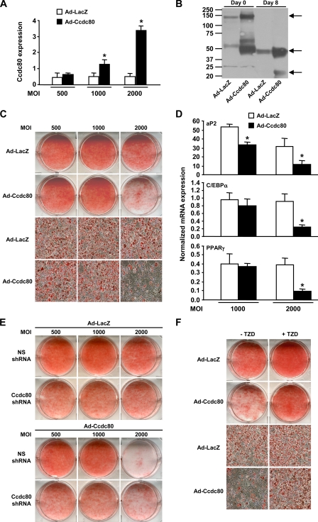
FIGURE 8.
Overexpression of Ccdc80 impairs adipogenesis. 3T3-L1 cells were infected with adenovirus encoding either LacZ (Ad-LacZ, white bars) or mouse Ccdc80 (Ad-Ccdc80, black bar) at various multiplicity of infection (m.o.i.). A, expression of Ccdc80 was determined by real-time PCR. B, secretion of Ccdc80. Conditioned medium from postconfluent (Day 0) and fully differentiated (Day 8) 3T3-L1 infected with adenovirus at a m.o.i. of 2000 was analyzed by Western blotting using an antibody raised against amino acids 840-854 of Ccdc80. Arrows indicate the full-length (∼140 kDa) and cleaved fragments (∼50 and ∼22 kDa) of Ccdc80 in conditioned medium from postconfluent (Day 0) and fully differentiated (Day 8) adipocytes. C, lipid accumulation. D, expression of adipogenic markers. Expression of aP2, C/EBPα, and PPARγ was determined by real-time PCR. E, lipid accumulation in 3T3-L1 cell lines expressing a NS shRNA or Ccdc80 shRNA infected with adenovirus encoding either LacZ or Ccdc80. F, effect of TZD on adipogenesis. Postconfluent (Day 0) 3T3-L1 cells infected with adenovirus at an m.o.i. of 2000 were differentiated with adipogenic inducers (dexamethasone, IBMX, and insulin) in the presence or absence of rosiglitazone (100 nm). C, E, and F, lipid accumulation was visualized by oil red O staining. Macroscopic and microscopic (100× magnification) images are shown. *, p < 0.05 versus Ad-LacZ.

