Figure 4.
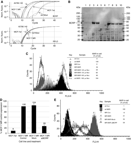
Analysis of BCRP expression and functionality in MCF-7.MR cells. (A) Expression of BCRP mRNA in MCF-7wt and A2780wt cells, and in the derivatives, MCF-7.MR and A2780.140: RT–PCR analysis of mRNA from untreated cells using Taqman expression assays (Applied Biosystems) containing primers and probes for BCRP, and for an endogenous control gene, RPLO. (B) Expression of BCRP protein in MCF-7wt and A2780wt cells, and in the derivatives, MCF-7.MR and A2780.140: protein from untreated cells was analysed by immunoblot as in Figure 3A. Lanes 1 and 2: MCF-7wt; lanes 3 and 4: MCF-7.MR; lanes 5 and 6: A2780wt; lanes 7 and 8: A2780.140; lanes 9 and 10: A2780.140 (no STX140 treatment for 8 weeks). Markers=MagicMark (Invitrogen). (C) The effect of Nov on the accumulation of MXR in MCF-7wt and MCF-7.MR cells: cells pre-treated with Nov were treated with 10 μM MXR +/− Nov, harvested by trypsinisation and resuspended in ice-cold PBS with 2.5% fetal calf serum. MXR accumulation was analysed using a flow cytometer (FACScan; Becton Dickinson) as in Figure 3C (wt=MCF-7wt; MR=MCF-7.MR; representative of two separate experiments). (D) Expression of BCRP mRNA in MCF-7.MR cells after BCRP siRNA transfection: RT–PCR analysis of mRNA from MCF-7wt and MCF-7.MR cells 48 h after transfection with 3 μg BCRP siRNA (Ambion, UK) using Taqman expression assays for BCRP, and for an endogenous control gene, RPLO. (E) The effect of BCRP siRNA transfection on the accumulation of MXR in MCF-7.MR cells: 48 h after transfection with 3 μg BCRP siRNA (Ambion, UK), MCF-7wt and MCF-7.MR cells were treated with 10 μM MXR +/− Nov, harvested by trypsinisation and resuspended in ice-cold PBS with 2.5% fetal calf serum. MXR accumulation was analysed using a flow cytometer (FACScan; Becton Dickinson) as in Figure 3C (wt=MCF-7wt; MR=MCF-7.MR; representative of two separate experiments).

