Figure 1.
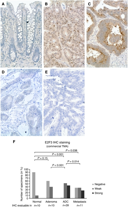
E2F3 expression detected by immunohistochemistry. E2F3 IHC analysis of the commercial TMA, COCA 912-5-OL, containing normal mucosa as well as benign and malignant colorectal specimens demonstrated that although E2F3 was not expressed by normal epithelial cells (A) E2F3 was found de novo synthesised by the majority of the investigated neoplastic tissues (B and C). The subcellular localisation of the de novo synthesised E2F3 protein was in some tumours found to be primarily nuclear (B) and cytoplasmic in others (C). Adenocarcinomas with no E2F3 staining (negative) were also observed, though only rarely (D). These were very similar to the ‘no primary’ antibody negative control (E). The frequency and intensity of E2F3 protein expression (combining nuclear and cytoplasmic staining) in adenoma, adenocarcinoma and metastasis samples were significantly higher than in normal mucosa (F). The same was the case when nuclear and cytoplasmic staining was evaluated individually (data not shown). The E2F3 IHC staining was evaluable and scored in 70 of the 71 tissue cores in the commercial TMA. P-values correspond to Fisher's exact tests. ADC=adenocarcinoma. All images are × 20. Staining: brown, E2F3; blue, haematoxylin counterstain.

