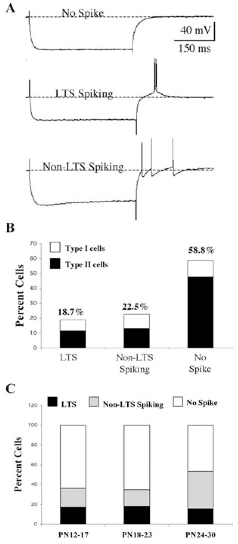
Fig. 6. Developmental changes in the rebound firing patterns displayed by Pf cells.

(A) Examples of the three types of rebound responses seen in Pf cells: No spike (top); low-threshold spike (LTS)-like spiking (middle); and non-LTS spiking (bottom). (B) Distribution of Type I and Type II cells with respect to the rebound response pattern. (C) Developmental changes in rebound response pattern plotted as a function of age group. LTS (filled bars), non-LTS spiking (shaded bars) and no spike (open bars). Note developmental decrease in no spike cells.
