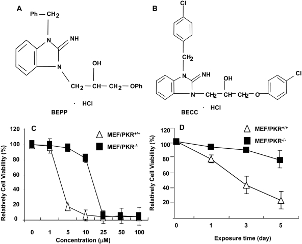
Fig. 1.
Chemical structure of BEPP (A) and its analog BECC (B). Effects of BEPP on proliferation of MEF/PKR(+/+) and MEF/PKR(-/-) cell lines. Cells were treated with various concentrations of BEPP for 72 h (C) or with 2.5 μM BEPP at the indicated time points (D). Cell viability was determined by using the SRB assay. Cells treated with DMSO were used as a control, with their viability set at 100%. Each data point represents the mean ± S.D. of three independent experiments.

