
Fig. 3.
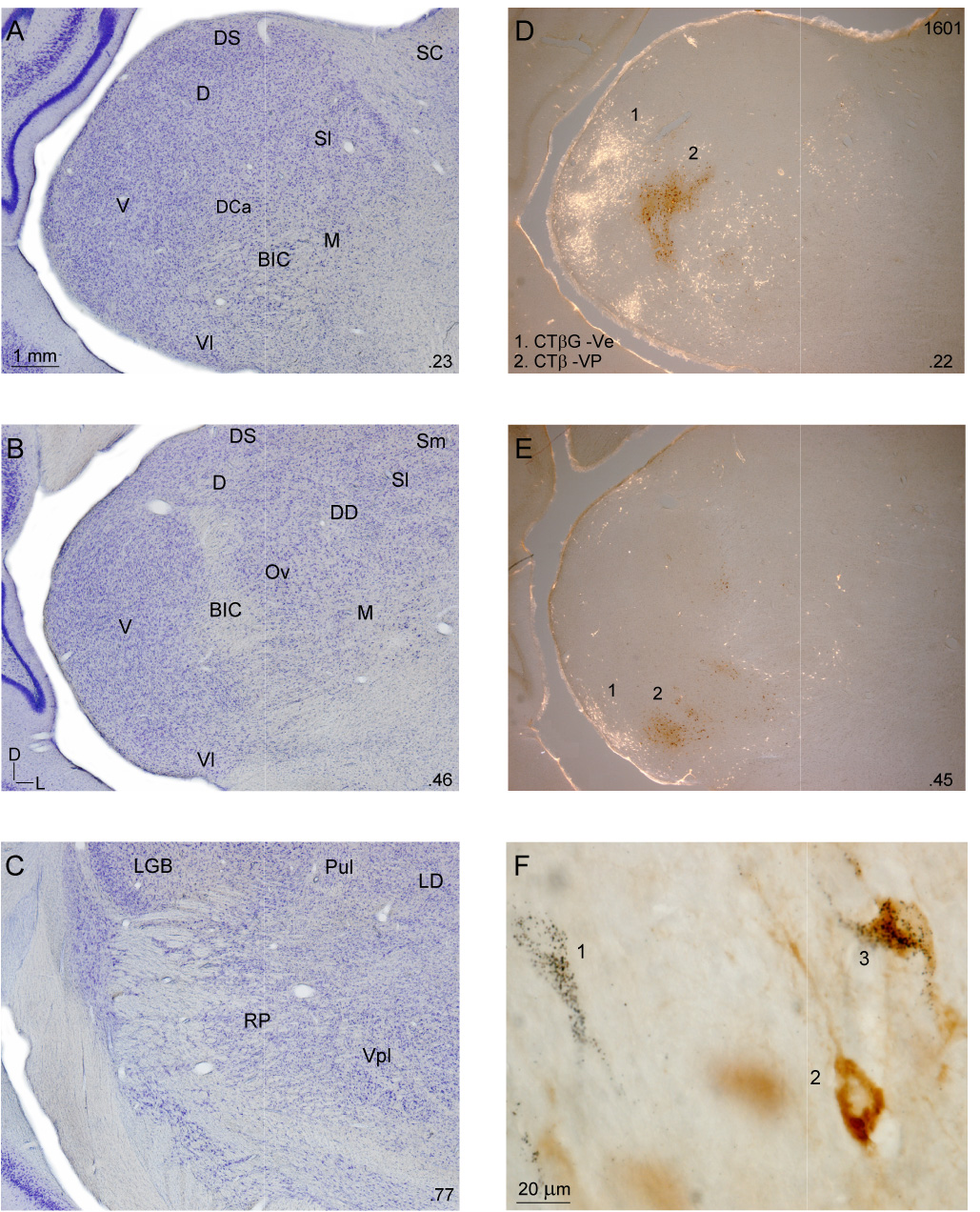
Thalamic cytoarchitecture and representative retrograde thalamic labeling. A–C: Nissl preparations at three caudorostral levels. Decimals (lower right), percent distance from the caudal tip of the MGB. A: Major divisions were present at 23% from the caudal tip. The pars lateralis of the ventral division (V) was conspicuous, with a characteristic laminar organization of dorsoventrally oriented neurons. The dorsal nucleus (D) had a lateral-to-medial arrangement and more densely packed neurons than the other dorsal division nuclei (the dorsocaudal (DCa), dorsal superficial (DS), ventrolateral (Vl), and lateral suprageniculate (Sl) nuclei). The dorsocaudal nucleus was receding by this level, while the dorsal superficial and ventrolateral nuclei had more dispersed cells and extended rostrally to ~70%. The lateral suprageniculate (Sl) had much larger neurons than other dorsal division nuclei and these are second in size to medial division (M) cells, which are sparser and form much of the MGB medial wall. B: Midway through the MGB, the brachium of the inferior colliculus (BIC) often divides the ventral division, with the pars ovoidea (Ov) medial to the brachium, and its laminar arrangement disrupted by it. The medial suprageniculate nucleus (Sm) extends from Sl to the dorsomedial thalamic border. C: At 77% from the caudal tip, the visual (LGN, Pul) and somatosensory (Vpl) thalamic nuclei border the MGB rostral pole. D–E: Representative retrograde labeling in bright-and darkfield illumination after injections in (1) ventral (Ve; CTβG) and (2) ventroposterior (VP; CTβ) areas. CTβG labeling (1: white cells). CTβ labeling (2: red-brown). Injections in VP and Ve labeled topographically segregated clusters in the ventral division (V), with lesser labeling in the dorsal nuclei and the medial division. F: CTβG (1), CTβ (2), and double-labeled (3) cells were readily distinguished.