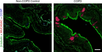
Figure 1.
Histologic evidence of mucous cell metaplasia in COPD. Representative photomicrographs of airway sections from patients with COPD and from control patients without COPD using whole lung explants obtained at the time of lung transplantation. Sections were immunostained for β-tubulin (green), MUC5AC (red), and CCSP (blue) and imaged by laser scanning confocal microscopy as described previously (29, 84). Bar = 20 μm.

