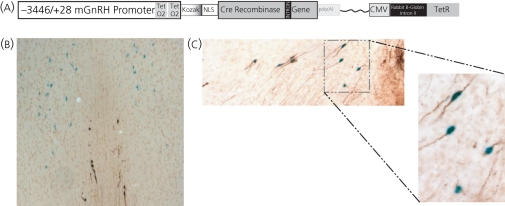
Fig.4.
Gonadotrophin-releasing hormone (GnRH) neurones exhibit CRE recombinase activity in doxycycline treated GnRH-CRETeR mice. (a) The construct used to produce the GnRH-CRETeR mouse is shown. A −3446 bp fragment of the mouse GnRH promoter was fused upstream of CRE recombinase as for the GnRH-CRE mouse. The proximal region of the GnRH promoter was modified to insert two Tet operators (TetO2). In series, the transgene also contains the CMV promoter regulating the Tet repressor gene. The rabbit β-globulin intron II is inserted between the promoter and the gene to enhance expression of the TetR gene. (b) Coronal sections from a bitransgenic GnRH-CRETeR/ROSALacZ mouse treated with Dox. Section includes the medial and lateral septum and the DBB. Tissue was treated with X-gal to visualise β-galactosidase activity and is seen as blue. GnRH neurones were stained as above and are brown. A population of X-gal stained neurones are seen in the lateral septum and do not express GnRH. Nine GnRH expressing neurones are seen in the medial septum and DBB, eight of which colocalised with X-gal staining. (c) GnRH neurones at the level of the organum vasculosum of the lamina terminalis/preoptic area at × 100 magnification. Ten GnRH immunostained neurones are depicted and nine co-stain blue indicating CRE recombinase activity. The boxed region is further displayed at × 250 magnification.

