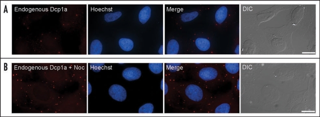
Figure 1.
Endogenous PBs were stained with an anti-Dcp1a antibody (red). The nucleus was counterstained with Hoescht (blue) and cells were also imaged in DIC (grey). (A) Normal distribution of PB in human cells. (B) Disruption of the microtubule network using nocodazole (noc) led to an increase in PB numbers. (Bar, 20 µm).

