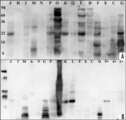
Figure 1.
Western blot analysis of CJD, disease and healthy controls. Urine samples of patient and control samples underwent proteolytic digestion with two different PK concentration and incubation time and were probed with anti-IgG-HRP anitbody. (A) Enriched urine samples from sCJD, vCJD, disease and healthy controls were treated with proteinase K (concentration 40 µg/ml for 20 min). Samples ID labelled with letters correspond to Table 1. After electrophoresis, protein was transferred to a nylon membrane using Bio-Rad instrument and probed with anti-IgG-HRP followed with ECL Plus addition. A Kodak Imager was used for photographic documentation. (B) Same or other samples were treated with proteinase K (concentration 60 µg/ml for 30 min). Samples ID labelled with letters correspond to Table 1, healthy control samples are shown with numbers. Protein was transferred via Bio-Rad and membrane probed with anti-IgG-HRP followed ECL Plus addition. Documentation was as above. (The exposure time for documentation was 5 minutes for A and B).

