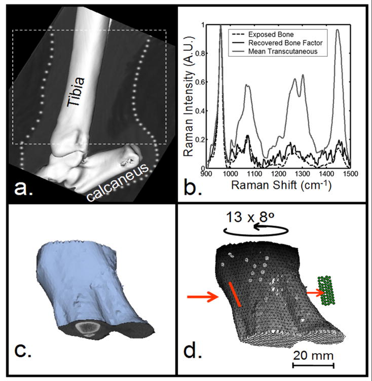
Fig. 1.
Development of mesh for tomographic reconstruction. a) Micro-CT image of canine hind limb section. b) Raman spectra of limb section. c) Geometric image of tissues from micro-CT data. d) Volumetric mesh created from (c). Input coordinates for reconstruction: tomographic projections and their associated scores. Illumination line (red) and collection fibers (green).

