
Figure 1.
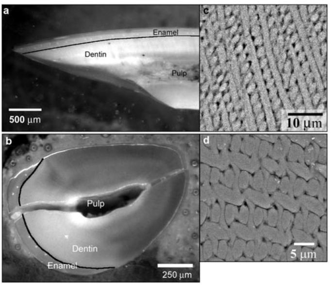
Optical micrographs of rat incisors cut in the mid-sagittal (a) and transverse (b) planes, which are normal to each other; black lines outline the dentino-enamel junction (DEJ). SEM-BSE micrographs of enamel polished in the mid-sagittal (c) and transverse (d) planes illustrate the difference in the enamel rod organization in these 2 planes. Note that the crack in (b) is a consequence of specimen preparation. Since the samples were not embedded, but rather were mounted in resin, the pulp cavity remained unfilled; this led to macrocracking of the sample during polishing. However, this crack did not affect the enamel integrity at the microscopic level.