
Figure 3.
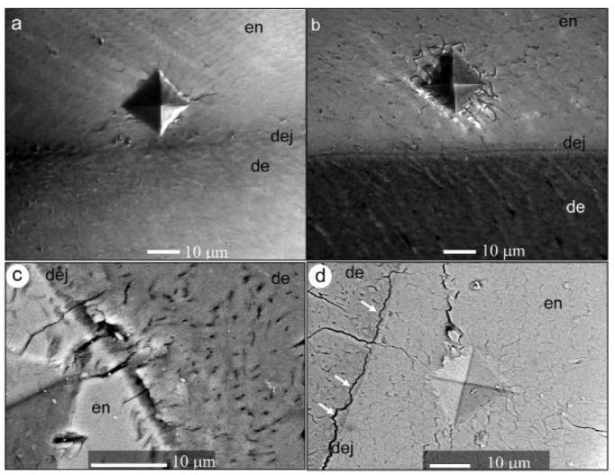
Optical micrographs representing characteristic damage produced by the microindenter in dry-untreated (a) and dry-treated (b) samples in the mid-sagittal plane. SEM-BSE micrographs demonstrating indentation damage in dry-treated samples in the mid-sagittal plane (c), and dry-treated samples in the transverse plane (arrows indicate areas of dentin attached to enamel) (d)