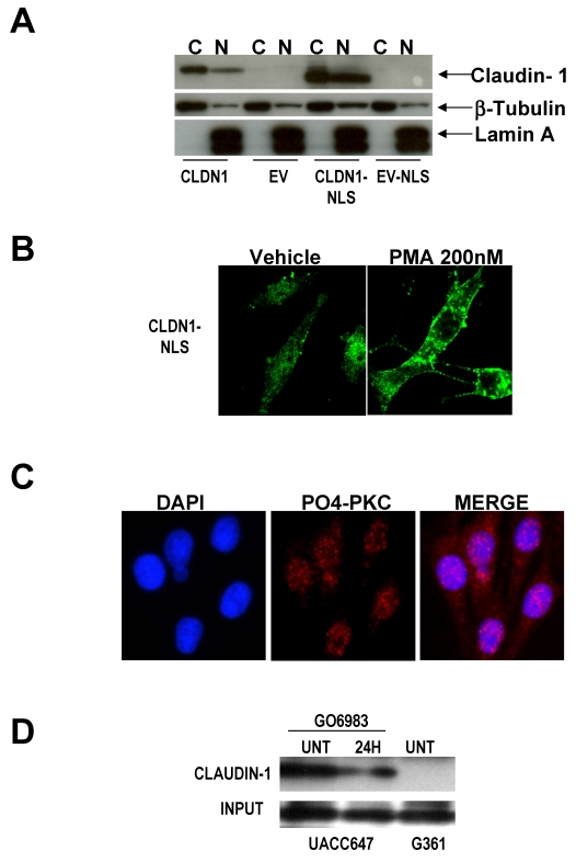
Figure 1.
Claudin-1 is not expressed solely in the nucleus even when under the direction of a nuclear localization signal. A) Cytoplasmic and nuclear extracts of G361 melanoma cells (low claudin-1 expressers) were transfected with wild type CLDN1 (CLDN1). By Western analysis, these transfectants demonstrate the presence of claudin-1 protein in the cytoplasm, with small amounts in the nucleus. When cells are transfected with CLDN1 under the control of a nuclear localization signal (CLDN1-NLS), there is increased expression of claudin-1 in the nucleus, but there are still large amounts of claudin-1 in the cytoplasm as well. B) All of the claudin-1 in the nucleus can be shuttled into the cytoplasm by treatment with the PKC activator PMA (phorbol ester). C) This implies that active PKC may exist in the nucleus of melanoma cells, so cells were stained with an antibody to pan-PO4-PKC. Confocal microscopy demonstrates that active PKC can be found in the nucleus of G361 melanoma cells. D) To determine if claudin-1 was a potential PKC substrate, cell lysates from claudin-1 high UACC647 cells, and claudin-1 low G361 cells were subjected to immunoprecipitation using a PKC substrate antibody, and western analysis for claudin-1 was performed. GO6983, a PKC inhibitor, decreases the amount of claudin-1 that is immunoprecipitated by the PKC substrate antibody. G361 cells have very little claudin-1 and thus, none is precipitated by the PKC substrate antibody, indicating the specificity of this immunoprecipitation.

