Abstract
Background
Tendinopathy of the flexor carpi ulnaris tendon is a rare entity. Recent research revealed the role of a neurovascular ingrowth at the point of pain in various tendinopathic locations, such as at the Achilles and patellar tendon, in plantar fasciitis as well as in supraspinatus and tennis elbow tendinopathy. However, beyond the elbow no such neovascularisation has been reported to date.
Methods
We present a 35‐year old tennis player suffering tremendous pain (visual analogue scale (VAS) rating of 9/10) at the flexor carpi ulnaris tendon with adjacent calcification in close proximity to the pisiform bone. The patient was assessed with power Doppler and laser Doppler quantification of neovascularisation at the point of pain.
Results
Power Doppler and laser Doppler quantification of neovascularisation at the point of pain identified higher capillary blood flow at three points over the painful vs the non‐painful tendon (146/240/232rU vs 93/74/70rU at the non‐affected side). Sclerosing therapy using polidocanol under power and laser Doppler guidance was initiated, with immediate decrease of capillary blood flow by 25% with resolution of the neovascularisation in power Doppler. Immediately following sclerosing, the patient's reported pain level on the VAS was reduced from 9/10 to 4/10. Following a short period of rest, eccentric training of the forearm muscle was initiated over 12 weeks with functional complete recovery and complete resolution of wrist pain.
Conclusion
Sclerosing therapy using polidocanol under power‐ and laser‐Doppler guidance can decrease capillary blood flow by 25% with resolution of the neovascularisation. Subsequent eccentric training of the forearm muscle over 12 weeks can result in complete resolution of wrist pain.
Pain on the ulnar side of the wrist is a common complain especially among tennis players.6 However, tendinopathy of the flexor carpi ulnaris (FCU) is a rare entity with reports published on case series basis only, while tendinopathy of the extensor carpi ulnaris tendon affects 1–2% of the population, most commonly in the fourth and fifth decade.3,6 A report from the Baylor College reported on five patients suffering FCU tendinopathy who failed to respond to conservative treatment with FCU debridement and histological evaluation, with degenerative tendinosis with angiofibroblastic hyperplastia in all specimens.1
Calcification secondary to tendon degeneration is encountered in the supraspinatus tendon in particular9 with enchondral ossification of the tendon being important for this condition.2 In various locations neurovascular infiltration is encountered in tendinopathy of the Achilles, the patellar and the supraspinatus tendon, as well as in tennis elbow tendinopathy. However, currently no data are available on the role of neovascularisation in forearm and wrist tendinopathy.4,5,7,8,10
What is already known on this topic
Neovascularisation plays an important role in tendinopathy in tennis elbow, supraspinatus tendinopathy, jumpers knee and Achilles pain.
Daily painful eccentric training over 12 weeks can reduce pain in Achilles mid‐portion tendinopathy as well as in patellar tendinopathy.
What this study adds
Sclerosing therapy using polidocanol under power‐ and laser‐Doppler guidance is feasible in flexor carpi ulnaris tendinopathy at the wrist with increased capillary blood flow, with immediate decrease of capillary blood flow by 25% with resolution of the neovascularisation in power‐Doppler.
Eccentric training of the forearm muscle over 12 weeks can result in complete resolution of wrist pain.
Case report
A 35‐year old tennis player was admitted to our department with severe pain on the ulnar aspect of his left wrist. Prior to admission, he experienced an episode of forearm pain related to exercise a year prior to admission. He complained of severe pain rating 9/10 on a visual analogue scale (VAS) even at gentle palpation over the flexor carpi ulnaris tendon. Due to the prior episode of forearm pain, rheumatological examination found slightly elevated CCP‐antibodies specific for rheumatoid arthritis (21 U/ml, normal <7 U/ml) with C‐reactive protein, IgA, IgG, IgM, C3, C4 and ANA within normal ranges.
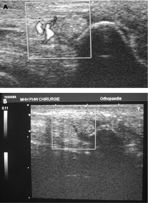
Plain radiographs of his wrist demonstrated a calcification in close proximity to the pisiform bone in the flexor carpi radialis tendon. Sonography verified the area of calcification to be in close proximity of the pisiform bone with hypoechogenic structure within the flexor carpi radialis tendon. Power Doppler (Toshiba Nemio 20 ultrasound system, Toshiba, London, UK) revealed a significant neovascularisation in the distal tendon proximal to the tendon calcification (fig 1A). Laser Doppler flowmetry incorporated in the Oxygen‐to‐see system (LEA Medizintechnik, Giessen, Germany) revealed an increased capillary blood flow at 8 mm tissue depths for three distinct positions along the distal flexor carpi radialis tendon at 1 cm apart, in contrast to the healthy contralateral side (from distal to proximal: 146/240/232rU at the symptomatic side vs 93/74/70rU at the asymptomatic side). Tendon oxygen saturation was slightly elevated at the corresponding three symptomatic vs the asymptomatic positions (81%/79%/88% vs 66%/65%/67%). However, postcapillary venous filling pressures at the FCU tendon were within the same range (46/39/50rU vs 38/55/41rU).
Figure 1 Neovascularisation of the flexor carpi radialis tendon determined with a power Doppler with an adjunct calcification in close proximity to the pisiform bone before (A) and following sclerosing (B) with 0.25% polidocanol using strict power Doppler control.
Using combined power Doppler and laser Doppler spectrophotometry a targeted sclerosing therapy was performed using 0.25% polidocanol in 0.1 ml titration volumes at the area of neovascularisation until resolution of the flow signal using the power Doppler (fig 1B), which was achieved with 1.5 ml of polidocanol. Capillary blood flow using the Oxygen‐to‐see system declined immediately at all three positions in the flexor carpi radialis tendon (113rU vs 146rU (distal), 219rU vs 240rU (middle), and 175rU vs 232rU (proximal). Tendon oxygenation was slightly elevated following sclerosing, from 81%/79%/88% to 97%/99%/91%, associated with slightly elevated (20%) postcapillary venous filling pressures. Pain immediately following the initial sclerosing decreased to 4/10 on the VAS, with further resolution of pain to VAS 0/10 following a 12 week interval of daily eccentric training of the forearm muscles, using the Thera‐Band® Flex‐Bar® (Thera‐Band, Hadamar, Germany) for eccentric training with 6×15 repetitions per day.
Discussion
Neovascularisation is evident beyond the elbow at the wrist level, such as in flexor carpi ulnaris tendinopathy. Power Doppler ultrasound associated with quantitative combined laser Doppler and spectrophotometry were capable of detecting the area of neovascularisation in flexor carpi ulnaris tendinopathy, where selective sclerosing therapy using polidocanol was performed under strict power Doppler control. Since the first sclerosing report at the Achilles tendon level,7 successful sclerosing has been demonstrated in patellar4 and elbow tendinopathy.10 Immediately following sclerosing, the patient's pain level was reduced by over 50% with further reduction within the next 2 weeks. Painful eccentric forearm training was initiated for a substantial and sustained modification of the wrist tendons.
Abbreviations
FCU - flexor carpi ulnaris
LD - laser Doppler
PD - power Doppler
VAS - visual analogue scale
Footnotes
Competing interests: None declared.
References
- 1.Budoff J E, Kraushaar B S, Ayala G. Flexor carpi ulnaris tendinopathy. J Hand Surg [Am] 200530125–129. [DOI] [PubMed] [Google Scholar]
- 2.Fenwick S, Harral R, Hackney R.et al Enchondral ossification in Achilles and patella tendinopathy. Rheumatology 200241474–476. [DOI] [PubMed] [Google Scholar]
- 3.Gabel G T. Acute and chronic tendinopathies of the elbow. Curr Opin Rheumatol 199911138–143. [DOI] [PubMed] [Google Scholar]
- 4.Hoksrud A, Ohberg L, Alfredson H.et al Ultrasound‐guided sclerosis of neovessels in painful chronic patellar tendinopathy: a randomized controlled trial. Am J Sports Med 2006341738–1746. [DOI] [PubMed] [Google Scholar]
- 5.Lind B, Ohberg L, Alfredson H. Sclerosing polidocanol injections in mid‐portion Achilles tendinosis: remaining good clinical results and decreased tendon thickness at 2‐year follow‐up. Knee Surg Sports Traumatol Arthrosc 2006141327–1332. [DOI] [PubMed] [Google Scholar]
- 6.Montalvan B, Parier J, Brasseur J L.et al Extensor carpi ulnaris injuries in tennis players: a study of 28 cases. Br J Sports Med 200640424–429. [DOI] [PMC free article] [PubMed] [Google Scholar]
- 7.Ohberg L, Alfredson H. Ultrasound guided sclerosis of neovessels in painful chronic Achilles tendinosis: pilot study of a new treatment. Br J Sports Med 200236173–177. [DOI] [PMC free article] [PubMed] [Google Scholar]
- 8.Rees J D, Wilson A M, Wolman R L. Current concepts in the management of tendon disorders. Rheumatology 200645508–521. [DOI] [PubMed] [Google Scholar]
- 9.Urist M R, Moss M J, Adams J M., Jr Calcification of the tendon. A triphasic local mechanism. Acta Pathol 196477594–608. [PubMed] [Google Scholar]
- 10.Zeisig E, Ohberg L, Alfredson H. Sclerosing polidocanol injections in chronic painful tennis elbow‐promising results in a pilot study. Knee Surg Sports Traumatol Arthrosc 2006141218–1224. [DOI] [PubMed] [Google Scholar]


