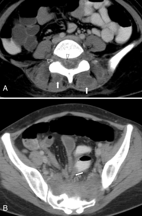
Figure 2).
A Axial computed tomography scan at the level of L5 to S1 disc space, showing multiple bilateral paraspinal abscesses (arrow) and ‘ring enhancing’ inflammatory masses in the epidural, and both right and left sacral foramina (open arrowheads). B Axial computed tomography scan of the lower pelvis, showing a thickened distal ileum tethered toward the inflammatory presacral abscess (arrow)

