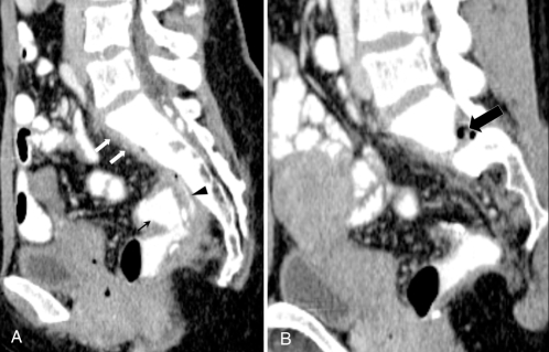
Figure 3).
A Sagittal computed tomography scan of the ileal pouch (black arrow), showing a leak from the posterior aspect of the ileoanal pouch (black arrowhead), with a thin layer of presacral abscesses (white arrows). B Sagittal computed tomography scan image to the left of the midline, showing pockets of gas with an ill-defined abscess in the intervertebral foramen in the epidural space (arrow)

