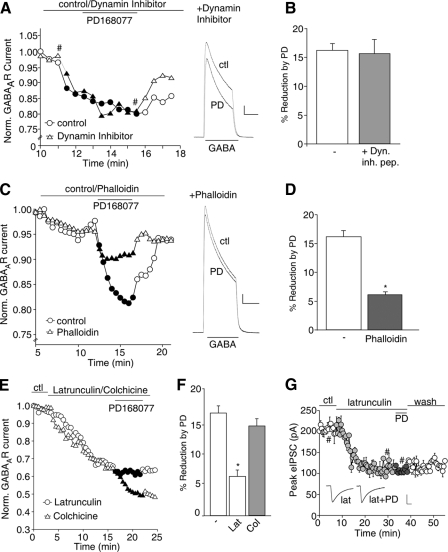
FIGURE 2.
D4 reduction of GABAAR currents is through an actin-dependent mechanism. A, plot of normalized peak GABAAR current as a function of time and PD168077 (30 μm) application in neurons dialyzed with or without the dynamin inhibitory peptide (50 μm). Inset, representative current traces (at time points denoted by #). Scale bar, 500 pA, 1 s. B, cumulative data (means ± S.E.) showing the percentage of reduction of GABAAR current by PD168077 in a sample of neurons in the absence (control) or presence of the dynamin inhibitory peptide. C, plot of normalized peak GABAAR current as a function of time and PD168077 application in neurons dialyzed with the actin stabilizer phalloidin (12.5 μm). Inset, representative current traces (at time points denoted by #). Scale bar, 500 pA, 1 s. D, cumulative data (mean ± S.E.) showing the percent reduction of GABAAR current by PD168077 in the absence or presence of phalloidin in a sample of neurons tested. *, p < 0.005, ANOVA. E, plot of normalized peak GABAAR current as a function of time and PD168077 application in neurons dialyzed with the actin depolymerizer latrunculin B (5 μm) or perfused with the microtubule depolymerizer colchicine (30 μm). F, cumulative data (mean ± S.E.) showing the percent reduction of GABAAR current by PD168077 in the absence or presence of latrunculin B or colchicine in a sample of neurons tested. *, p < 0.005, ANOVA. G, plot of evoked IPSC as a function of time and PD168077 application in a PFC slice perfused with latrunculin B. Inset, representative IPSC traces (at time points denoted by #). Scale bar, 50 pA, 20 ms. ctl, control; PD, PD168077; Dyn. inh. pep., dynamin inhibitory peptide; Lat, latrunculin B; Col, colchicine.

