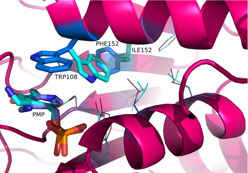
FIGURE 3.
Modeling of the active site of AGT-Ma and F152I variant in the PMP form. The putative location of Trp108 at the active site of AGT-Ma and F152I variant in the PMP form is illustrated. Trp108, Phe152, and PMP are represented as blue sticks in AGT-Ma, whereas Trp108, Ile152, and PMP are represented as cyano sticks in F152I variant. Oxygen atoms are colored red, nitrogen atoms blue, and phosphorus orange. This figure was rendered using PyMOL (17).

