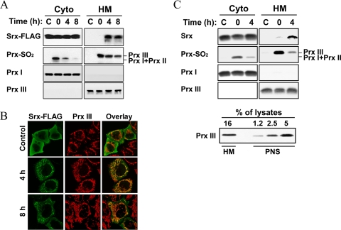
FIGURE 2.
Mitochondrial translocation of Srx under oxidative conditions. HeLa cells transfected with an expression vector for FLAG-tagged Srx (A and B) and normally grown A549 cells (C) were exposed to 200 μm H2O2 for 10 min and then allowed to recover from oxidative stress as in Fig. 1. A and upper panel of C, after the indicated times, cell homogenates were separated into nuclear pellet and postnuclear supernatant (PNS). 0.5 ml of PNS was further separated into 0.5 ml of cytosolic (Cyto) and mitochondria-enriched heavy membrane (HM) fractions, and then the latter was prepared as 0.05 ml of lysates. Equal volumes (0.03 ml) of Cyto fractions and HM lysates were analyzed by immunoblotting for the indicated proteins. Lower panel of C, the indicated percent volumes of aliquots from HM lysates (0.05 ml) or PNS (0.5 ml) were subjected to immunoblot analysis for Prx III. B, cells were stained for the FLAG epitope (green) and Prx III (red) and then examined by confocal microscopy.

