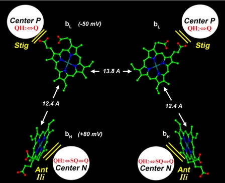
FIGURE 1.
Location of QH2/Q binding sites and b hemes in the yeast bc1 complex dimer. Edge-to-edge distances between heme tetra-pyrrole rings are indicated by arrows along with the heme redox midpoint potentials as measured in the isolated yeast bc1 complex. The approximate locations of center P and center N are also shown along with the redox reactions occurring in each site and their respective inhibitors: stigmatellin (Stig), antimycin (Ant), and ilicicolin (Ili). The structure was constructed from coordinates of the yeast bc1 complex, Protein Data Bank (PDB) code 1EZV (3).

