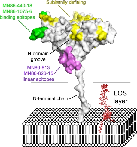
FIGURE 6.
Proposed model representing LP2086-B01 in the bacterial membrane. The flexible linker between residues 1 and 19 allows the protein to span the LOS layer of the outer membrane. Subfamily-defining residues (yellow) populate one region on one face of the protein oriented toward the extracellular space. In violet, the linear epitopes for the monoclonals MN86-626-15 and MN86-813 are oriented toward the membrane surface and are not accessible by these mAbs in flow cytometry experiments. In green, the binding epitopes for the monoclonals MN86-440-18 and MN86-1075-6 are also exposed to the extracellular space. A single LOS molecule is indicated in red in the model. The carbohydrate torsional angles have been manually set to achieve maximum extension of the headgroup.

