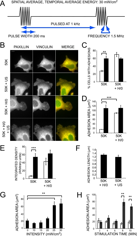
FIGURE 1.
Ultrasound stimulates focal adhesion formation independently of syndecan-4 engagement. A, schematic representation of the ultrasound wave form. B, wild-type MEFs were spread on 50K for 2 h before stimulating with syndecan-4 ligand (H/0) for 40 min either with (closed bars) or without (open bars) ultrasound for 20 min. Fixed cells were stained for paxillin (green) and vinculin (red). Bar, 10 μm. C, 400 cells/condition were scored for focal adhesion formation. Total focal adhesion area (D) or total focal adhesion-integrated density (E) of 25-35 cells/condition was measured using image J software. F, average focal adhesion length of 20 cells/condition. G, focal adhesion area of MEFs stimulated with a range of ultrasound intensities. H, focal adhesion area of MEFs stimulated with ultrasound for a range of durations. Error bars, S.E. Significance values are as follows: *, p < 0.05; **, p < 0.01; ***, p < 0.001. Images and analyses are representative of experiments performed on at least three separate occasions.

