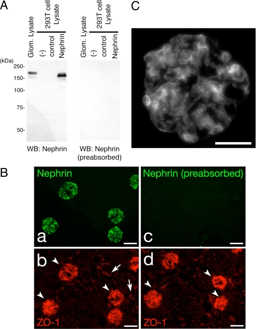
FIGURE 1.
Detection of Nephrin with the anti-Nephrin antibody. A, lysates from isolated rat glomeruli, untransfected HEK293T cells, and HEK293T cells transiently transfected with plasmid encoding Nephrin or a control vector were separated on SDS-PAGE (10%), transferred to nitrocellulose membrane, and immunoblotted with anti-Nephrin (left) or anti-Nephrin preabsorbed with the peptide used for immunization (right). B, adult rat cryosections were analyzed by indirect immunofluorescence microscopy (magnification, ×100) after incubating sections simultaneously with anti-zonula occludens (ZO-1) and rabbit Nephrin antibody (a and b) or anti-Nephrin antibody preabsorbed with the peptide used for immunization (c and d) to confirm Nephrin staining in glomeruli (arrowheads) but not in the tubular cells (arrows). Scale bars, 100 μm. C, nephrin staining shows a typical podocyte pattern along the glomerular capillary loops. Magnification, ×400; scale bars, 50 μm.

